How to identify an implant when nobody told you the brand
A screw loosens ten years later and your dentist asks which system it is. If you do not know, it is recoverable more often than people expect — here is the order to work through.
What this article establishes
- Start with the treating clinic; many keep records for years and will send them on request
- A periapical radiograph often narrows the system down, because connections differ visibly
- Ask for the record before you fly home, not after — it is one piece of paper
The situation
Something has gone wrong with an implant you have had for years. A crown has come loose, a screw has fractured, or the gum around it is unhappy. You go to a dentist near where you live, and the first question is: which implant is it?
If you know, this is a routine repair. If you do not, it becomes a problem — and for patients treated abroad without proper records, it is the most predictable long-term difficulty in the whole of implant dentistry.
The good news is that it is recoverable far more often than people assume. Here is the order to work through.
Why the brand alone is not enough
First, calibrate what you are actually looking for, because “Nobel Biocare” is not an answer.
Manufacturers produce many systems, and systems change over time. NobelActive, NobelParallel, NobelReplace and N1 do not share the same connection geometry, and within each there are platform diameters that are not interchangeable. A component that fits one will not seat correctly in another.
So what a dentist needs is the system name plus the platform diameter, and ideally the length and the exact position. “It is a Nobel” narrows the search usefully and does not end it.
Step 1 — Ask the clinic that treated you
Obvious, frequently skipped, and successful more often than people expect.
Many clinics retain treatment records for years, including clinics abroad, and will send them on request. Email in writing, give your full name, date of birth and the approximate treatment date, and ask specifically for:
- The implant system name
- The diameter and length of each implant
- The tooth positions
- The abutment and restorative components used
- The implant labels from the packaging, if they were kept
Ask for photographs of the labels rather than a typed summary if you can — the sticker carries the exact reference and removes any transcription error.
If the clinic has closed, ask whether records were transferred. In many jurisdictions there are obligations around retention, and successor practices sometimes hold the archive.
Step 2 — Take a radiograph
If the paperwork is gone, the implant itself carries evidence.
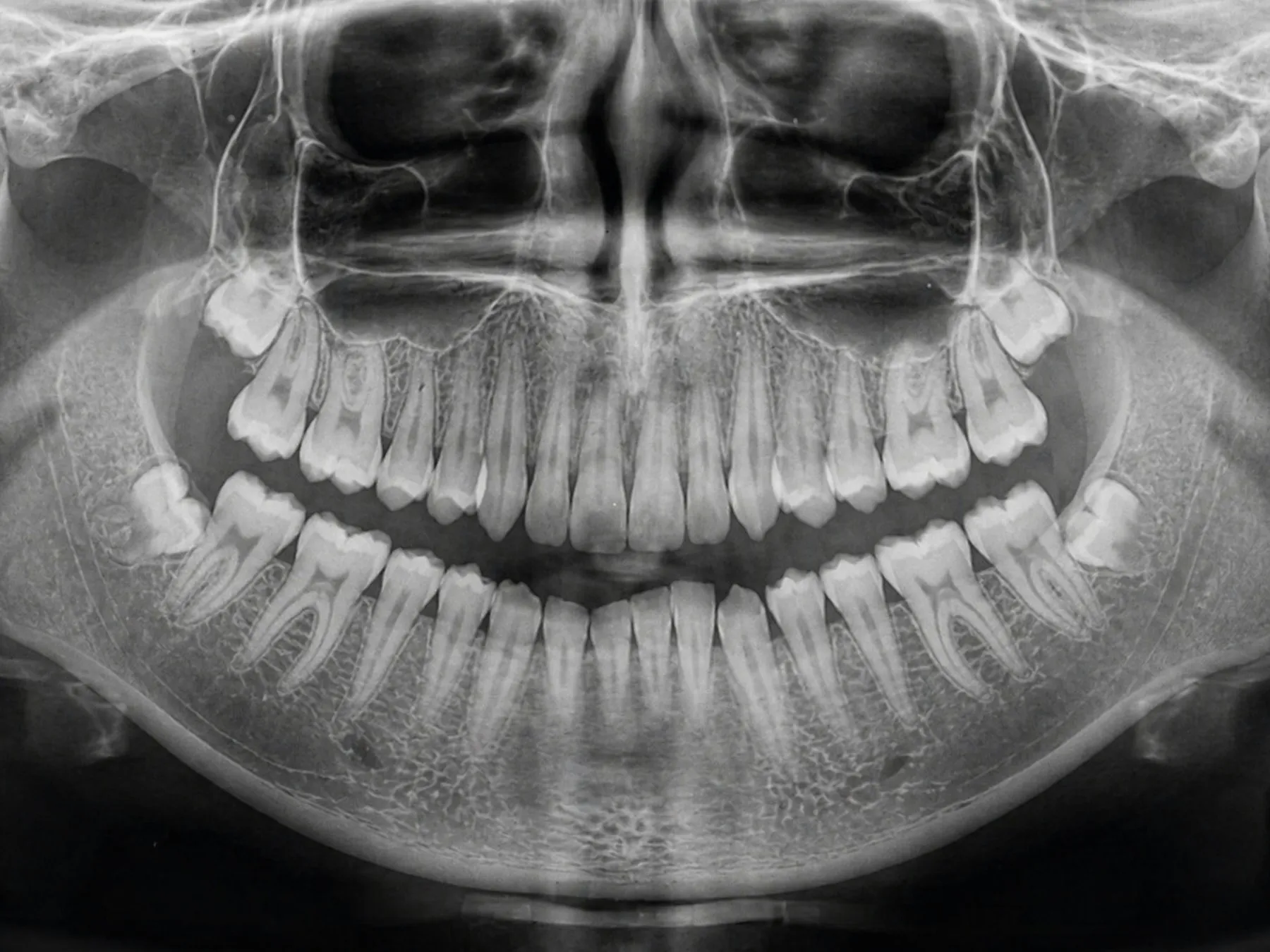
A periapical radiograph — a small, focused X-ray of that tooth — taken by any dentist will show the implant’s outline: thread pattern, body shape, and crucially the geometry of the connection at the top. These differ visibly between manufacturers, and specialists compare the image against identification atlases and databases built for exactly this purpose.
Identification from an image is not guaranteed. It frequently gets close enough to source a compatible component, and occasionally it produces a definitive answer. It costs one appointment and it is the obvious second move.
There is a public reference for this too: independent implant guides publish identification resources and manufacturer profiles which a dentist can work against.
Step 3 — Look at the connection directly
If the crown is already loose or coming off, the connection underneath can be examined and measured directly. That is usually conclusive.
A clinician will not remove sound work purely to look — the risk is not worth it — but where the restoration is failing anyway, the diagnostic information comes free with the repair.
Step 4 — Consider what actually needs to happen
Worth saying plainly: sometimes exact identification stops mattering.
If the implant itself is sound and only the crown has fractured, the fix may be a new crown on the existing abutment, which sidesteps the question. If the implant has failed and is coming out, its identity is largely historical. Full identification matters most in the middle case — implant fine, component broken, replacement part required.
Ask your dentist which of the three you are in before spending appointments on identification you may not need.
What a dentist is actually doing when they identify one
Understanding the method helps you judge how confident an answer is.
The identifiable features on a radiograph are the thread profile (spacing, depth and shape), the body outline (parallel or tapered, and how sharply), the apex — the tip, which differs markedly between manufacturers — and above all the connection geometry at the top, where the abutment seats. That last feature is the most distinctive, because connections are the part manufacturers design to be proprietary.
A clinician compares those features against reference collections built for the purpose. Some are published atlases; some are software tools that narrow candidates from an uploaded image.
The result is usually a shortlist rather than a single name — “this is one of three systems, all of which take this component” is frequently enough to proceed. Where it is not, direct examination of the connection settles it.
What defeats identification is a poor-quality image. A radiograph taken at an angle foreshortens the implant and distorts exactly the features being compared. If a first attempt is inconclusive, a repeat taken with a proper holder is a reasonable next step before concluding it cannot be done.
Why this problem is worse for patients treated abroad
Not because clinics abroad are worse — because of distance and time.
If you were treated near home and something goes wrong, you return to the practice that did the work, and the records are in the building. If you were treated in another country five years ago, you are relying on an email to a clinic that may have changed premises, staff or ownership, in a language you may not share, about a treatment they performed once.
None of that is insurmountable and much of it is avoidable. It simply means the record matters considerably more for an international patient than a domestic one, and it is the reason this article exists on a site aimed at people who travel for treatment.
It is also a fair question to put to any clinic before you book: if I need a component in ten years and cannot reach you, what will I have that lets someone else help me?
What to insist on so this never happens
If you are being treated now, this section is the one that saves you the other four.
Before you leave — before you fly home, if you are travelling — ask for your implant record in writing. It should state:
- Manufacturer and system name
- Diameter and length of every implant placed
- Tooth position for each, in standard notation
- Date of placement
- Treating clinic and clinician
- Abutment and restorative components used
- The product labels from the packaging
- Follow-up and maintenance recommendations
Photograph it and store the photograph somewhere you will still have access to in ten years — email it to yourself, put it in whatever cloud storage you actually use. A printed card in a drawer is worth less than it looks.
This is standard practice at a clinic that expects you to be somebody else’s patient for maintenance, which — if you were treated abroad — you will be. It is why Taki Dent issues the record as part of finishing treatment rather than on request, and it is a fair thing to ask any clinic whether they do the same before you commit.
What it costs to identify one
Worth setting expectations, because patients sometimes delay this over an imagined bill.
A periapical radiograph is among the least costly procedures in dentistry — the sort of image taken routinely at a check-up. The identification work itself is usually a short part of the appointment rather than a separate service, and most dentists will do it while assessing the problem you came in with.
Cost appears only if identification proves genuinely difficult and a specialist opinion is needed, or if a component has to be ordered on a best-guess basis and turns out not to seat. Both are uncommon, and both are cheaper than the alternative.
Set against that: an unidentifiable implant that ends up being removed and redone because no matching part could be found is one of the more expensive outcomes in implant dentistry. The economics of spending one appointment on identification are not close.
What this says about choosing a clinic
Treat the answer to “what documentation will I leave with?” as a screening question.
A clinic that answers immediately and specifically is a clinic that has thought about your next ten years. A clinic that is vague, or that offers a certificate with a brand logo and no dimensions, has told you something useful about how it sees the relationship.
Implants are not maintenance-free. They need cleaning, review and occasionally repair, and most of that will happen near where you live rather than where they were placed. The record is what connects the two.
If you are stuck now
Send whatever you do have — the radiograph, an old invoice, the clinic’s name, even just the approximate year — with a photograph of the area concerned. Partial information is worth substantially more than none: a date and a country narrow the field of candidate systems considerably.
The clinical team at Taki Dent can tell you whether the implant appears identifiable from what you have, what imaging would settle it, and whether identification is even the thing standing between you and the repair. A final diagnosis and treatment plan can only be confirmed after clinical examination and appropriate radiological assessment — but working out what you are dealing with can usually start from an image.